Rydberg Quantum Enhanced Tracker (QET).
A project with a collaboration between William & Mary and Thomas Jefferson National Lab in Newport News. This project is a direct application of Rydberg electric field sensing. An electron beam provides a DC electric field gradient that can be detected by Rydberg atoms.
 When Rydberg atoms are subject to a uniform DC electric field, the EIT transmission peak created from optical detection will have a frequency shift proportional to the strength of the electric field, and the polarizability of the atomic state. The Rydberg QET project aims to use the shift of the transmission peak to measure electron beam current and give dimenstions of beam width and position.
Fluorescence Based Rydberg Measurements
To spatially map the electric fields, it is convenient to use a camera to construct a spatial map of electric fields across the cell volume. In a collaborative effort with NIST we have been working on doing fluorescence based measurements of a probe laser in an atomic volume to map highly spatially varying fields. Here we show a cartoon overview of our detection scheme where a small number of atoms are around an electron beam. The electric field generated by the electron beam can be derived through Gauss's law as just a wire of varing levels of enclosed charge. With laser beams that are thinner than the electron beam itself we can get a cross section of the electric field created by the electron beam. We do this by using a camera to take a video of the probe laser in the cell.
 Through this method we have shown that we can reconstruct values of electric field along the sensing volume. The figure here shows how we can reconstruct a value of electric field at each position the camera sees. With this information we can fit the reconstructed electric field to extract parameters of the elctron beam such as current, position and width.
Transmission Based Rydberg Measurements
As an alternative to the fluorescence based mehtod, we can also look at the transmisison of the probe laser through the cell. Instead of the shifts to the resonance, there is a field gradient, so we must look at distortions to the EIT peak that are presented as braodening rather than relative shifts. To do this we pulse the electron beam, and see how the field changes to avoid unwanted charging effects and have a clean signal due to only the presence of the electron beam.
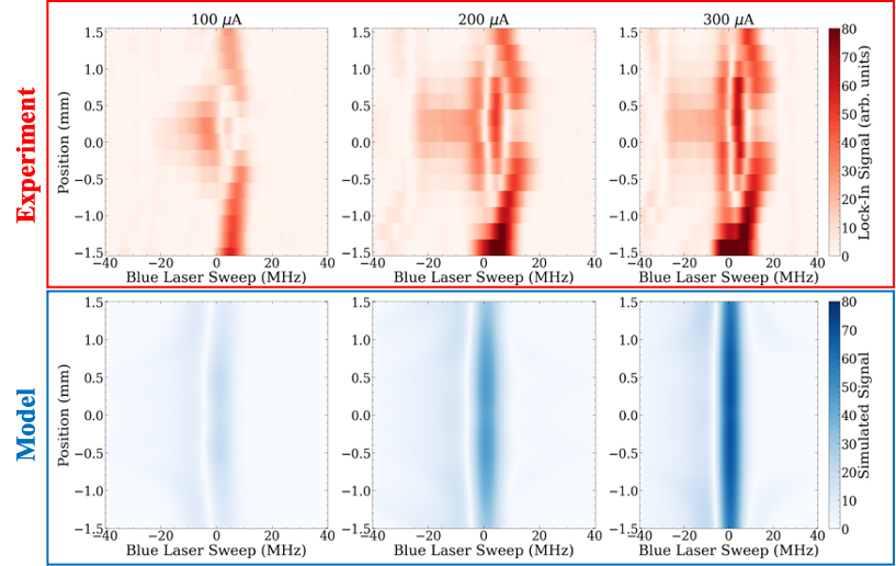 Using this method have found a spatially dependent signal that makes the EIT respond in a similar way to how a simplified model shows the peak should be distorted. Pulsing the electron beam has mitigated much of the charging effects that have been problematic, but we still are sensitive to a spatial electric gradient not due to the electron beam within our sensing cell.
To map the spatial gradient we have attempted moving the electron beam around the lasers, moving the lasers around the electron beam, and also angling the laser beams and moving the overlapped-crossed beam region around the cell.
Using this method have found a spatially dependent signal that makes the EIT respond in a similar way to how a simplified model shows the peak should be distorted. Pulsing the electron beam has mitigated much of the charging effects that have been problematic, but we still are sensitive to a spatial electric gradient not due to the electron beam within our sensing cell.
To map the spatial gradient we have attempted moving the electron beam around the lasers, moving the lasers around the electron beam, and also angling the laser beams and moving the overlapped-crossed beam region around the cell.